Sri Aurobindo Medical College & Postgraduate Institute, Indore (M.P.)
DEPARTMENT OF CARDIAC SCIENCES
ABOUT THE DEPARTMENT
The Department of Cardiac Sciences at Sri Aurobindo Medical College & PG Institute, Indore, is one of the best ”Center for Excellence” for Cardio vascular interventions & Surgeries. From its inception since 2011, more than 67000 patients have been treated successfully. We are providing comprehensive cardiac services from preventive cardiology to interventional of complex cardiovascular ailments. We have two state of art Cath labs, very well equipped with all modern softwares and applications. The Department is offering academic courses in the campus with the constant efforts and guidance of our visionary Founder Chairman Dr. Vinod Bhandari. We are running DM Course in cardiology with 3 intake capacity.
INFRASTRUCTURE – STATE OF
ART TECHNOLOGY
· Cath Lab:
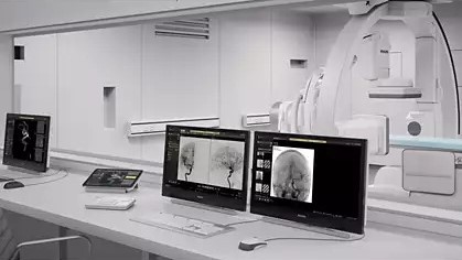
Philips Azurion 7 B20/15 Top
End Cath Lab System

Welcome to the Future of Advance
cardiac care & interventional procedures
We are proud to announce the
major addition of our new cardiovascular X-ray interventional suite. This is a
significant investment in promoting the delivery of enhanced patient care
Using the latest X-ray
imaging technology, our team of specialists can now perform a wider range of
minimally invasive diagnostic and interventional cardiovascular procedures in a
patient-focused environment.
At the heart of our new interventional suite is the Philips
Azurion imaging system. This advanced technology helps to optimize patient
comfort, and enables us to provide an advanced level of care for your patients.
Features
·
Latest high resolution 20-inch detector to produce excellent
Image quality
·
State-of-the-art hardware platform and software architecture
·
Azurion platform is designed to perform advance
interventional procedures
·
It brings the innovative workflow to reduce procedure time in
the lab
·
Lowest radiation exposure in the industry
·
Software packages that provide high resolution Dynamic
coronary Roadmap and Stentboost
live for Cardiac and
3DRA, Live 3D Roadmap and XperCT- CT like soft tissue
imaging for neuro and vascular which will be used for
enhanced diagnosis, planning and treatment
GE IGS 320 – Premium 12” Flat Panel “Low
Dose” Digital Cath Lab
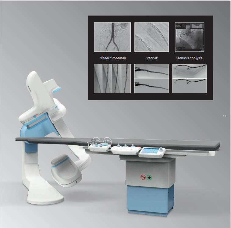
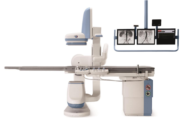
The IGS 320
is designed to sustain long, demanding, and complex procedures.
FEATURES
ð
High resolution imaging at low dose
The entire image chain is optimized, from the digital
detector, collimator, tube, and exposure management strategy to image processing,
so that we can easily view small details as well as low-contrast objects during
both fluoroscopy and record imaging
ð
The proven platform for our complex cardiac interventions
With its
20,5 x 20,5 cm detector, the IGS 320 is an optimal choice for cardiovascular
procedures, allowing us to reach steep angulations, and cover the whole heart.
With its offset C -arm we can benefit from increased coverage.
We can also perform long vascular exams by rotating the C-Arm on both sides. In
addition to being the perfect platform to support high procedure volumes, the
IGS 320 was designed to address a wide variety of complex procedures.
·
Well Equipped ICU (16 Bedded including 10 Ventilator Beds)
·
Philips and Wipro GE Color Doppler and Echo Systems
·
Unimed Tread Mill
·
Philips HOLTER Unit
·
Wipro GE Ambulatory BP System
SERVICES / FACILITIES
·
Coronary Angiography
·
Coronary Angioplasty
·
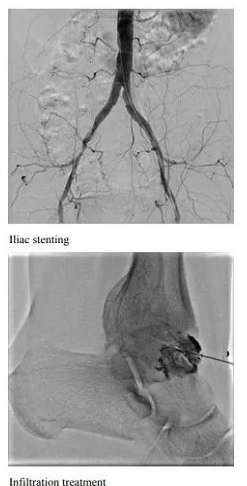
Peripheral Angiography
·
Peripheral Angioplasty
·
Pacemaker Implantation(Single & Dual)
·
CRT-D
·
TAVI
·
EVAR
·
Electro-Physiology & Radio Frequency Ablation (EP Study
& RFA)
·
IVUS
·
Rota-ablation
·
OCT
·
Valve Ballooning
·
Device Closure
·
Echo – Cardiography (Transthoracic
& Trans-esophageal)
·
Dobutamine Stress Echocardiography
·
TMT
·
HOLTER
·
Stress Imaging (Spect, PET, Cardiac
MRI)
·
All Pediatric Procedures (ASD, VSD, PDA ligation etc.)
·
Coronary Artery Bypass Surgery (CABG)
·
MidCab (Minimal Access ByPass
Surgery)
·
Valve Surgeries – Valve repair and Valve Replacement
·
Peripheral Graft Surgeries
·
Emboelectomies
·
Congenital Heart Defect Surgeries
·
Thoracotomy
·
PeriCardiactomy
·
Aortic Aneurysm Surgeries
ACADEMIC
COURSES
·
DM Cardiology Course (Running with 3 Seats)
Proposed Courses:
·
Fellowship in Clinical Cardiology – (3 Seats, 2 Years, post MBBS)
·
ECG & TMT Technician Course – (3 Seats, 2 Years, 12th
Science & Basic Computer knowledge)
·
Echocardiography Technician Course – (3 Seats, 2 Years, 12th
Science & Basic Computer knowledge)
·
Cath Lab Technician Course – (3 Seats, 2 Years, 12th
Science & Basic Computer knowledge)
FACULTIES
|
DEPARTMENT OF CARDIAC
SCIENCES |
||
|
Sr. No. |
Names |
Designation |
|
1 |
Dr.
Pradeep Kumar Gupta
|
Professor
& Head (Cardiology) |
|
2 |
Dr.
Rakesh Jain |
Assoc.
Professor (Cardiology) |
|
3 |
Dr.
Gourav Singh |
Assoc.
Professor (Cardiology) |
|
4 |
Dr. Ram Rawat |
Assoc.
Professor (CTVS) |
|
5 |
Dr.
Suyash Tated |
Asst.
Professor (Cardiology) |
|
6 |
Dr.
Harshal Pamecha |
Asst.
Professor (Cardiology) |
|
7 |
Dr. Nishith Bhargava |
Asst.
Professor (CTVS) |
uMI Vista Digital
160 Slice PET/CT System
Procedures :
·
Whole Body Scan
·
Organ Scans
·
High Dose Therapy
·
CT Angio
Clinical
Benefits :
Complete Digital PET-CT solution with the highest accuracy of subcentimeter lesion detection
·
Faster whole body scan in 4 beds
·
Complete Patient scan within 5-6
min
·
50 % Lower patient dose
·
High definition images for
detailed information
·
Sub centimetre
lesion detectability
·
One stop cardiac imaging solution
with 160 slice Cardiac CT
·
Automatic quality control system
to monitor the performance of the system
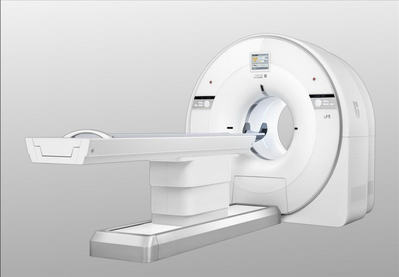
160 Slice uMI Vista Digital PET / CT System
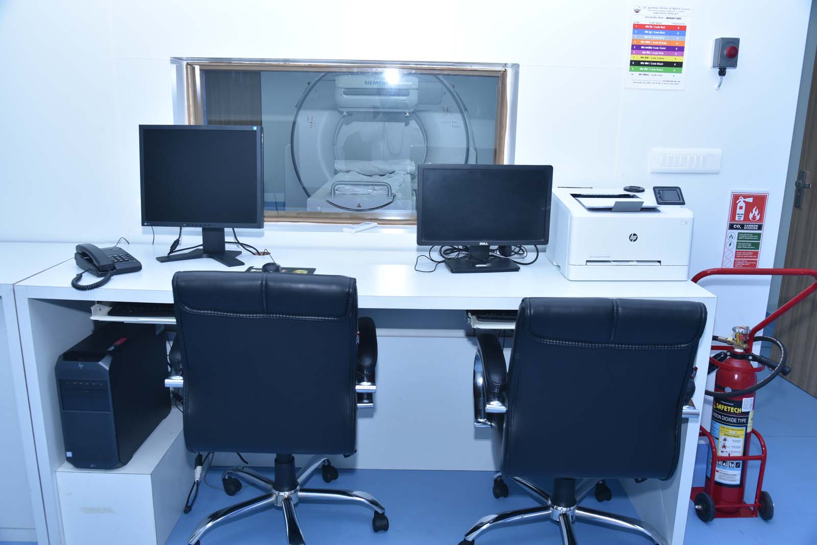
SYMBIU
EVO EXCEL DUAL HEAD SPECT GAMMA CAMERA
This camera enables very high quality functional images in half
the time for most of the procedures as there are two camera heads scanning the
patient
simultaneously.
SPECT's
ability-to reveal physiologic and metabolic change makes it a unique tool for diagnosing
and treating various diseases. Shorter photomultiplier tubes combined with lean
front-end electronics reduce analog noise and improve performance and the wide
variety of collimators are optimized to enable high sensitivity, low septa1
penetration and high resolution even when the detectors are farther from the
patient resulting in best quality images.
Nuclear
Medicine studies performed on SPECT camera at our facility are
:
1. SKELETAL SCINTIGRAPHY - WHOLEBODY BONE
SCAN, THREE PHASE BONE SCAN
2. ENDOCRINE SYSTEM - THYROID
SCINTIGRAPHY, RADIO IODINE THYROID/WHOLE BODY IMAGING, PARATHYROID IMAGING, 131L-
MLBG ADRENOMEDULLARY IMAGING ETC.
3. HEPATOBILIARY SYSTEM - LIVER / SPLEEN
IMAGING, HLDA HEPATOBILIARY SCAN
4. GENITOURINARY SYSTEM - STATIC RENAL
IMAGING, DYNAMIC RENOGRAM STUDIES, VUR STUDIES, TESTICULAR PERFUSION SCAN
5. GASTROINTESTINAL SYSTEM- GI BLEED,
GASTRIC EMPTYING STUDIES, GASTRO ESOPHAGEAL REFLUX IMAGING, MECKEL'S
DIVERTICULUM ETC
6. MYOCARDIAL
PERFUSION STUDIES
- PHYSIOLOGICAL / PHARMACOLOGICAL STRESS -REST SCAN, VIABILITY, RESTING MUGA
SCAN
7. BRAIN PERFUSION STUDIES - 99M
TC-ECD/STROKE
8. LUNG VENTILATION / PERFUSION STUDIES
9. LYMPHOSCINTIGRAPHY AND SENTINEL NODE
MAPPING
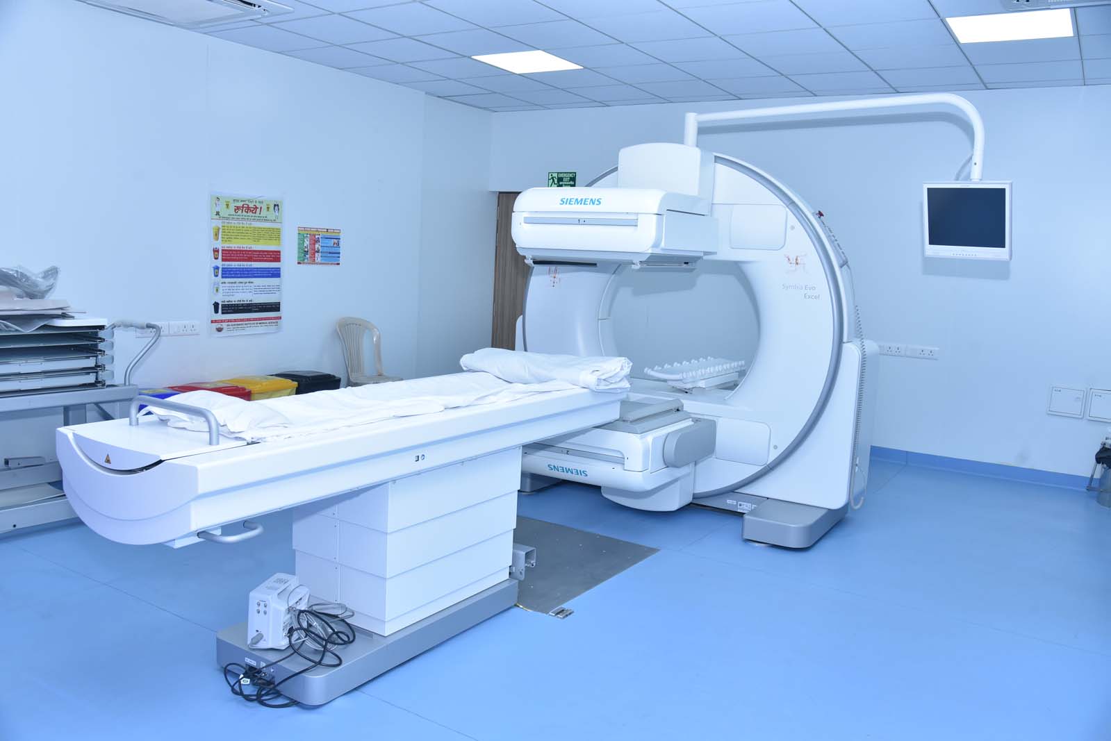
SYMBIU EVO EXCEL DUAL HEAD SPECT GAMMA CAMERA